Reduce Background. Preserve Signal. Improve Reproducibility.
MIBIslide® Blue is engineered to minimize slide-derived background while maintaining tissue integrity, enabling more consistent and reliable MIBI imaging results.
- Reduced slide-derived background signal
- Improved tissue-edge clarity and segmentation
- Consistent biomarker intensity across samples
- Designed for FFPE and frozen tissue sections, with seamless integration into H&E workflows

Designed for Workflow Continuity
Designed for FFPE and frozen tissue sections, with seamless integration into H&E workflows.
Standard Staining Compatibility
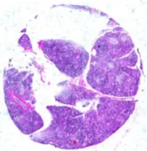
Standard H&E staining and digital slide scanning can be performed prior to MIBI staining and imaging on the same tissue section.
Pathology-Compatible Workflow
STANDARD PATHOLOGY
MIBI
H&E Staining
Digital Imaging
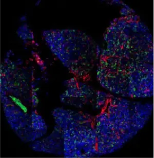
MIBI Staining
MIBI Imaging
Reproducible Signal Across Serial Sections
Matched segmentation parameters across serial sections demonstrate consistent expression distributions and reproducible single-cell measurements across experiments.
EXPRESSION DISTRIBUTION

Cell Object Area (microns)
SINGLE-CELL MEASUREMENTS

Workflow Continuity
Tissue sections move from routine pathology workflows into high-plex spatial proteomics without disruption, supporting continuity across discovery, translational, and longitudinal studies.
MIBIslide® Blue
Reduce technical variability. Focus on biological insight.
- Reduced Background Signal
- Compatibility with Standard Pathology Workflows
- Reproducible Single-Cell Measurements
Integrate MIBIslide® Blue into Your Spatial Proteomics Workflow
Never Miss an Update
Subscribe to our newsletter to get the latest news, tips, and updates delivered straight to your inbox.